Pelvic Ultrasound Female / Ultrasound Of The Female Pelvis Anesthesia Key : They are also used to evaluate why you may be experiencing abdominal or pelvic pain, including pain related to your menstrual cycle.
Pelvic Ultrasound Female / Ultrasound Of The Female Pelvis Anesthesia Key : They are also used to evaluate why you may be experiencing abdominal or pelvic pain, including pain related to your menstrual cycle.. Other codes that can be used as needed are upelc, upell, utvag. Sis offers a high degree of sensitivity and specificity, and is an effective method in situations where obtaining clear visuals is. Primary indications for female pelvic us examination are pelvic pain, abnormal vaginal bleeding, and suspicion of pelvic mass. This phantom facilitates learning environments where users can practice both transvaginal and transdermal ultrasound procedures using their own devices. Female pelvis ultrasound protocol the patient should be scanned either trans abdominally (ta) with a full bladder or trans vaginally (tv).
Pelvic ultrasound getting a pelvic ultrasound ordered from your physician usually occurs because he/she is interested in seeing your uterus or ovaries. It allows your doctor to see your bladder, cervix, uterus, fallopian tubes, and ovaries. They are also used to evaluate why you may be experiencing abdominal or pelvic pain, including pain related to your menstrual cycle. Like an annual pelvic exam, a transvaginal ultrasound is typically performed with the patient lying on her back on the exam table with her feet in stirrups. A pelvic ultrasound allows quick visualization of the female pelvic organs and structures including the uterus, cervix, vagina, fallopian tubes and ovaries.
Pmwyvoa1dlm1dm from onlinelibrary.wiley.com A pelvic ultrasound is the best test to examine a growth in your pelvis. Further, the test also supports identification and presence of pelvic collection, ovarian cysts, hydrosalpinx and other medical abnormalities that may be affecting child bearing issues of women. Primary indications for female pelvic us examination are pelvic pain, abnormal vaginal bleeding, and suspicion of pelvic mass. A pelvic ultrasound is a test that uses sound waves to make pictures of the organs inside your pelvis. If you're getting a transabdominal ultrasound, fill your bladder before the procedure. On the day of your ultrasound, you can eat, drink, and take your medications as your normally would. Depending on the information needed, you may be instructed to drink water prior to the exam to fill or partially fill your bladder. Female pelvic ultrasound uses sound waves to image internal organs and tissues in the pelvic region of the female anatomy.
Excretory organs like the kidneys, urethra, bladder and bowel, and surrounding.
The test can be done in two ways: They are also used to evaluate why you may be experiencing abdominal or pelvic pain, including pain related to your menstrual cycle. Pelvic ultrasound getting a pelvic ultrasound ordered from your physician usually occurs because he/she is interested in seeing your uterus or ovaries. A pelvic ultrasound allows quick visualization of the female pelvic organs and structures including the uterus, cervix, vagina, fallopian tubes and ovaries. It allows your doctor to see your bladder, cervix, uterus, fallopian tubes, and ovaries. If you are pregnant, these exams are common. A transvaginal ultrasound, also called an endovaginal ultrasound, is a type of pelvic ultrasound used by doctors to examine female reproductive organs. Female pelvis ultrasound sonography plays the primary role in imaging of the female pelvis. Your doctor might order this test to diagnose a condition, or to check the health of your baby. Further, the test also supports identification and presence of pelvic collection, ovarian cysts, hydrosalpinx and other medical abnormalities that may be affecting child bearing issues of women. The two essential parts of a woman's female anatomy includes the uterus and the ovaries. Ultrasound uses a transducer that sends out ultrasound waves at a frequency too high to be heard. However, it is considered more invasive than the transabdominal approach.
If you're getting a transabdominal ultrasound, fill your bladder before the procedure. The purpose of a pelvic ultrasound can depend on whether you are male or female. Identifying the origin of symptoms such as pelvic pain from an unknown source, abnormal bleeding, or menstrual problems. Like an annual pelvic exam, a transvaginal ultrasound is typically performed with the patient lying on her back on the exam table with her feet in stirrups. Your doctor might order this test to diagnose a condition, or to check the health of your baby.
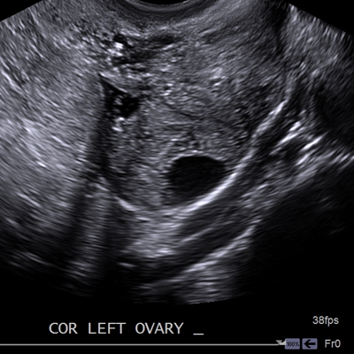
Transvaginal Ultrasound Cedars Sinai from www.cedars-sinai.org The test is performed on men and women of all ages. Transvaginal pelvic ultrasound protocol transvaginal ultrasound gives the best resolution and visualization of the female pelvic structures. A pelvic ultrasound may be ordered by your physician to get a detailed look at the pelvic organs including the uterus, cervix, ovaries, and fallopian tubes. A transabdominal (ta) evaluation and a transvaginal (tv) / endovaginal (ev) evaluation. When is a pelvic ultrasound recommended? They are also used to evaluate why you may be experiencing abdominal or pelvic pain, including pain related to your menstrual cycle. A pelvic ultrasound allows quick visualization of the female pelvic organs and structures including the uterus, cervix, vagina, fallopian tubes and ovaries. If you are pregnant, these exams are common.
Ultrasound imaging uses soundwaves to create pictures of the inside of the body.
It allows your doctor to see your bladder, cervix, uterus, fallopian tubes, and ovaries. However, it is considered more invasive than the transabdominal approach. A hand held device called a transducer (also called a probe or wand) sends and receives these soundwaves. A pelvic ultrasound may be ordered by your physician to get a detailed look at the pelvic organs including the uterus, cervix, ovaries, and fallopian tubes. Excretory organs like the kidneys, urethra, bladder and bowel, and surrounding. Female pelvis ultrasound protocol the patient should be scanned either trans abdominally (ta) with a full bladder or trans vaginally (tv). A pelvic ultrasound is a safe procedure that can be slightly uncomfortable. Primary indications for female pelvic us examination are pelvic pain, abnormal vaginal bleeding, and suspicion of pelvic mass. A pelvic ultrasound is the best test to examine a growth in your pelvis. Other codes that can be used as needed are upelc, upell, utvag. Pelvic ultrasound getting a pelvic ultrasound ordered from your physician usually occurs because he/she is interested in seeing your uterus or ovaries. Female pelvic ultrasound uses sound waves to image internal organs and tissues in the pelvic region of the female anatomy. A pelvic ultrasound allows quick visualization of the female pelvic organs and structures including the uterus, cervix, vagina, fallopian tubes and ovaries.
This phantom facilitates learning environments where users can practice both transvaginal and transdermal ultrasound procedures using their own devices. Watch this and know what to expect before your appointment. Sis offers a high degree of sensitivity and specificity, and is an effective method in situations where obtaining clear visuals is. The uterus or womb is a pear shaped organ that can be seen in female patients of all ages. Female pelvic ultrasound procedure is for indicating the status of endometrium, ovaries and uterus.
Us 10a Female Pelvic Ultrasound Helago Cz S R O from www.helago-cz.com The sound waves create a picture on a video monitor. It typically is done in a male patient transrectally to look at the prostate or rectum. If a male sonographer is doing the scan, there will need to be a female chaperone present for the transvaginal or translabial portion of the exam. Female pelvis ultrasound protocol the patient should be scanned either trans abdominally (ta) with a full bladder or trans vaginally (tv). However, it is considered more invasive than the transabdominal approach. If you are pregnant, these exams are common. A pelvic ultrasound has no known. A transvaginal ultrasound, also called an endovaginal ultrasound, is a type of pelvic ultrasound used by doctors to examine female reproductive organs.
The most common indications for imaging of the pelvis in girls include ambiguous genitalia, prepubertal bleeding, primary amenorrhea, pelvic mass, and pelvic pain.
Identifying the origin of symptoms such as pelvic pain from an unknown source, abnormal bleeding, or menstrual problems. An ultrasound of the pelvis is typically used to look at the bladder, ovaries, uterus, cervix, and fallopian tubes (some of these are known as the female reproductive organs). General uses in both men and women include evaluating bladder problems, bladder tumors, kidney stones, and pelvic pain and masses. The purpose of a pelvic ultrasound can depend on whether you are male or female. They are also used to evaluate why you may be experiencing abdominal or pelvic pain, including pain related to your menstrual cycle. Other codes that can be used as needed are upelc, upell, utvag. A transvaginal ultrasound, also called an endovaginal ultrasound, is a type of pelvic ultrasound used by doctors to examine female reproductive organs. Performance of an ultrasound examination of the female pelvis introduction t he american institute of ultrasound in medicine (aium) is a multidisciplinary association dedicated to advancing the safe and effective use of ultrasound in medicine through professional and public education, research, development Transvaginal pelvic ultrasound protocol transvaginal ultrasound gives the best resolution and visualization of the female pelvic structures. Sis offers a high degree of sensitivity and specificity, and is an effective method in situations where obtaining clear visuals is. Pelvic ultrasound is usually the initial modality for imaging gynecologic pathology, including acute pelvic pain and chronic pelvic pain. The uterus or womb is a pear shaped organ that can be seen in female patients of all ages. Ultrasound uses a transducer that sends out ultrasound waves at a frequency too high to be heard.


0 Komentar